
Research ArticleDermatologyImmunology
Open Access |  10.1172/jci.insight.205826
Substance P–NK1R signaling of keratinocytes initiates the skin inflammatory environment required for allergic contact dermatitis
Sumeet Manandhar,1 Mohna Bandyopadhyay,1 Olga A. Tkacheva,1 William J. Shufesky,2,3 Gregory A. Gibson,4 Simon C. Watkins,4,5,6 Adrian E. Morelli,2,3,5 and Adriana T. Larregina1,5,6
1Department of Dermatology,
2Thomas E. Starzl Transplantation Institute,
3Department of Surgery,
4Department of Cell Biology and Center for Biological Imaging, and
5Department of Immunology, University of Pittsburgh, School of Medicine, Pittsburgh, Pennsylvania, USA.
6McGowan Center for Regenerative Medicine, University of Pittsburgh, Pittsburgh, Pennsylvania, USA.
Address correspondence to: Adriana T. Larregina, Thomas E. Starzl Biomedical Science Tower, Room W1147, 200 Lothrop Street, Pittsburgh, Pennsylvania 15213, USA. Phone: 412.651.5545; Email: adrianal@pitt.edu.
Authorship note: SM and MB contributed equally to this work. ATL and AEM have been designated as co–senior authors.
Find articles by Manandhar, S. in: PubMed | Google Scholar
1Department of Dermatology,
2Thomas E. Starzl Transplantation Institute,
3Department of Surgery,
4Department of Cell Biology and Center for Biological Imaging, and
5Department of Immunology, University of Pittsburgh, School of Medicine, Pittsburgh, Pennsylvania, USA.
6McGowan Center for Regenerative Medicine, University of Pittsburgh, Pittsburgh, Pennsylvania, USA.
Address correspondence to: Adriana T. Larregina, Thomas E. Starzl Biomedical Science Tower, Room W1147, 200 Lothrop Street, Pittsburgh, Pennsylvania 15213, USA. Phone: 412.651.5545; Email: adrianal@pitt.edu.
Authorship note: SM and MB contributed equally to this work. ATL and AEM have been designated as co–senior authors.
Find articles by Bandyopadhyay, M. in: PubMed | Google Scholar
1Department of Dermatology,
2Thomas E. Starzl Transplantation Institute,
3Department of Surgery,
4Department of Cell Biology and Center for Biological Imaging, and
5Department of Immunology, University of Pittsburgh, School of Medicine, Pittsburgh, Pennsylvania, USA.
6McGowan Center for Regenerative Medicine, University of Pittsburgh, Pittsburgh, Pennsylvania, USA.
Address correspondence to: Adriana T. Larregina, Thomas E. Starzl Biomedical Science Tower, Room W1147, 200 Lothrop Street, Pittsburgh, Pennsylvania 15213, USA. Phone: 412.651.5545; Email: adrianal@pitt.edu.
Authorship note: SM and MB contributed equally to this work. ATL and AEM have been designated as co–senior authors.
Find articles by Tkacheva, O. in: PubMed | Google Scholar
1Department of Dermatology,
2Thomas E. Starzl Transplantation Institute,
3Department of Surgery,
4Department of Cell Biology and Center for Biological Imaging, and
5Department of Immunology, University of Pittsburgh, School of Medicine, Pittsburgh, Pennsylvania, USA.
6McGowan Center for Regenerative Medicine, University of Pittsburgh, Pittsburgh, Pennsylvania, USA.
Address correspondence to: Adriana T. Larregina, Thomas E. Starzl Biomedical Science Tower, Room W1147, 200 Lothrop Street, Pittsburgh, Pennsylvania 15213, USA. Phone: 412.651.5545; Email: adrianal@pitt.edu.
Authorship note: SM and MB contributed equally to this work. ATL and AEM have been designated as co–senior authors.
Find articles by Shufesky, W. in: PubMed | Google Scholar
1Department of Dermatology,
2Thomas E. Starzl Transplantation Institute,
3Department of Surgery,
4Department of Cell Biology and Center for Biological Imaging, and
5Department of Immunology, University of Pittsburgh, School of Medicine, Pittsburgh, Pennsylvania, USA.
6McGowan Center for Regenerative Medicine, University of Pittsburgh, Pittsburgh, Pennsylvania, USA.
Address correspondence to: Adriana T. Larregina, Thomas E. Starzl Biomedical Science Tower, Room W1147, 200 Lothrop Street, Pittsburgh, Pennsylvania 15213, USA. Phone: 412.651.5545; Email: adrianal@pitt.edu.
Authorship note: SM and MB contributed equally to this work. ATL and AEM have been designated as co–senior authors.
Find articles by Gibson, G. in: PubMed | Google Scholar
1Department of Dermatology,
2Thomas E. Starzl Transplantation Institute,
3Department of Surgery,
4Department of Cell Biology and Center for Biological Imaging, and
5Department of Immunology, University of Pittsburgh, School of Medicine, Pittsburgh, Pennsylvania, USA.
6McGowan Center for Regenerative Medicine, University of Pittsburgh, Pittsburgh, Pennsylvania, USA.
Address correspondence to: Adriana T. Larregina, Thomas E. Starzl Biomedical Science Tower, Room W1147, 200 Lothrop Street, Pittsburgh, Pennsylvania 15213, USA. Phone: 412.651.5545; Email: adrianal@pitt.edu.
Authorship note: SM and MB contributed equally to this work. ATL and AEM have been designated as co–senior authors.
Find articles by Watkins, S. in: PubMed | Google Scholar
1Department of Dermatology,
2Thomas E. Starzl Transplantation Institute,
3Department of Surgery,
4Department of Cell Biology and Center for Biological Imaging, and
5Department of Immunology, University of Pittsburgh, School of Medicine, Pittsburgh, Pennsylvania, USA.
6McGowan Center for Regenerative Medicine, University of Pittsburgh, Pittsburgh, Pennsylvania, USA.
Address correspondence to: Adriana T. Larregina, Thomas E. Starzl Biomedical Science Tower, Room W1147, 200 Lothrop Street, Pittsburgh, Pennsylvania 15213, USA. Phone: 412.651.5545; Email: adrianal@pitt.edu.
Authorship note: SM and MB contributed equally to this work. ATL and AEM have been designated as co–senior authors.
Find articles by Morelli, A. in: PubMed | Google Scholar
1Department of Dermatology,
2Thomas E. Starzl Transplantation Institute,
3Department of Surgery,
4Department of Cell Biology and Center for Biological Imaging, and
5Department of Immunology, University of Pittsburgh, School of Medicine, Pittsburgh, Pennsylvania, USA.
6McGowan Center for Regenerative Medicine, University of Pittsburgh, Pittsburgh, Pennsylvania, USA.
Address correspondence to: Adriana T. Larregina, Thomas E. Starzl Biomedical Science Tower, Room W1147, 200 Lothrop Street, Pittsburgh, Pennsylvania 15213, USA. Phone: 412.651.5545; Email: adrianal@pitt.edu.
Authorship note: SM and MB contributed equally to this work. ATL and AEM have been designated as co–senior authors.
Find articles by Larregina, A. in: PubMed | Google Scholar
Published September 1, 2026 - More info
JCI Insight. 2026;11(19):e205826. https://doi.org/10.1172/jci.insight.205826.
© 2026 Manandhar et al. This work is licensed under the Creative Commons Attribution 4.0 International License. To view a copy of this license, visit http://creativecommons.org/licenses/by/4.0/.
Received: February 13, 2026; Accepted: August 25, 2026


-
Results
The SP-NK1R axis is required for the skin innate immunity triggered by DNCB and croton oil. Signaling of the NK1R by SP potentiates the adaptive immune response of the CHS, the mouse equivalent of ACD, to strong contact sensitizers (6, 8, 18). However, the roles of SP or the NK1R at early stages of the CHS sensitization are not completely understood. Therefore, we tested whether the SP-NK1R axis is required for the skin innate immunity underlying the irritant effect of the contact sensitizer. This innate immune response is necessary for the efficient development of the adaptive immune response that accounts for the effector phase of the CHS (1, 3, 12, 18). To address the role of SP in the skin irritant effect of haptens, Tac1KO (lacking SP) or WT mice were sensitized on the right ear with topical application of DNCB (1% in 1:4 acetone/olive oil) or treated on the left ear with vehicle. For this experiment, the sensitization in the mouse ear was chosen because it allows monitoring of the innate immune response by measurement of the increase of ear thickness following application of the contact sensitizer, which cannot be done in mice sensitized in the abdominal skin. The increase of ear thickness was assessed 24, 48, and 72 hours after sensitization, and parameters indicative of local innate immunity were analyzed in tissue samples of ears at the peak of the inflammatory response to DNCB, 24 hours after sensitization (Supplemental Figure 1A; supplemental material available online with this article; https://doi.org/10.1172/jci.insight.205826DS1). As expected from the DNCB irritant effect, the thickness of the ear and the epidermis and dermis, and the density of skin-infiltrating leukocytes, increased significantly in WT mice sensitized with DNCB, and these parameters were significantly lower in equally treated Tac1KO mice lacking SP (Figure 1, A–D, and Supplemental Figure 1A).
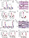 Figure 1
SP and the NK1R are necessary for the skin innate immunity triggered by topical exposure to DNCB. (A) Skin innate immune response to topical DNCB by percentage of ear thickness increase in WT and Tac1KO mice. (B) Microscopic images of ear sections of mice in A showing epidermal-dermal thickness (vertical lines) and infiltrating leukocytes. Scale bar: 50 μm. (C and D) Quantification of epidermal and dermal thickness (C) and infiltrating leukocytes (D) in skin sections shown in B. (E) Innate immune response to DNCB by percentage of ear thickness increase in WT, NK1RKO, and Mrgb2KO mice. (F) Microscopic images of ear sections of mice in E showing epidermal-dermal thickness (vertical lines) and infiltrating leukocytes. Scale bar: 50 μm. (G and H) Quantification of epidermal and dermal thickness (G) and infiltrating leukocytes (H) in the skin sections shown in F. In A and E, each circle represents 1 mouse (n = 5). B and F are images of samples obtained 24 hours after sensitization. H&E; original magnification, ×200. In C and G, each circle represents 1 measurement (n = 10). In D and H, each circle represents the quantification of skin leukocytes in 1 microscopic high-power field (HPF), ×500 (n = 20). Means ± 1 SD were analyzed by 1-way ANOVA and post hoc Student-Newman-Keuls test; *P < 0.05, **P < 0.01, ***P < 0.001, ****P < 0.0001.
To determine whether the proinflammatory effects of SP were mediated via the NK1R (high-affinity SP receptor) or Mas-related G protein–coupled receptor B2 (Mrgb2) (low-affinity SP receptor), NK1RKO, Mrgb2KO, and WT mice were skin-sensitized with DNCB (right ear) or treated with vehicle (left ear), and the parameters of local innate immunity induced by the hapten were analyzed in the ear skin. The ear and epidermal-dermal thickness and the density of skin-infiltrating leukocytes of DNCB-treated ears were significantly reduced in NK1RKO mice compared with the values detected in Mrgb2KO mice or WT controls, and there were no significant differences between WT and Mrgb2KO mice (Figure 1, E–H).
Next, we analyzed whether the SP-NK1R axis was relevant not only to the induction of the innate immune response of ACD caused by DNCB, but also to that induced by skin exposure to chemicals that trigger irritant contact dermatitis (ICD). For these experiments, WT, Tac1KO, and NK1RKO mice were treated in the dorsal skin of the right ear with croton oil (CO; 2% in acetone). Evaluation of ear thickness 2, 4, 6, 8, and 24 hours after application of CO in WT mice showed significant increase, which peaked from 4 to 6 hours after treatment (Supplemental Figure 1B) and correlated with intense dermal edema and skin-infiltrating polymorphonuclear leukocytes. These parameters were significantly diminished in Tac1KO and NK1RKO mice evaluated up to 24 hours after treatment (Supplemental Figure 1, C–G).
Thus, the SP-NK1R axis is essential to initiate the local innate immunity required for the sensitization phase of ACD and the development of ICD.
The SP-NK1R axis in keratinocytes is necessary for the proinflammatory skin environment and the adaptive immune response to DNCB. Keratinocytes, the most abundant cells of the skin that are exposed to haptens, express the NK1R and are in contact with SP-secreting sensory nerves (18–22, 24, 50, 51). Therefore, we analyzed the role of SP-NK1R signaling specifically in keratinocytes during the initiation of the cutaneous innate immune response triggered by DNCB. To address that, K14Cre/+ NK1RKO mice, with specific deletion of the NK1R in keratinocytes (18), and their littermate (Creneg) controls were treated topically with DNCB or vehicle on the dorsal skin of the right and left ear, respectively. Parameters indicative of development of an innate immune response were assessed in the ear skin 24 hours after sensitization. Compared with their littermate controls, K14Cre/+ NK1RKO mice exhibited significantly less increase of ear and epidermal-dermal thickness and of skin-infiltrating leukocytes in response to DNCB treatment (Figure 2, A–D). Consequently, compared with WT mice and their littermates, K14Cre/+ NK1RKO mice exhibited a significant decrease in the effector CHS response following skin sensitization and elicitation with DNCB, and this decrease was comparable to that observed in Tac1KO and global NK1RKO mice (Supplemental Figure 2, A–D).
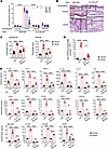 Figure 2
Expression of NK1R in keratinocytes is required for the skin inflammatory environment triggered by topical DNCB. (A) Skin innate immune response to DNCB in K14Cre/+ NK1RKO (K14-NK1RKO) mice and their littermate (Litt) controls assessed by percentage of ear thickness increase. (B) Microscopic images of the ears of mice in A showing epidermal-dermal thickness (vertical lines) and skin-infiltrating leukocytes. Scale bar: 50 μm. (C and D) Quantification of epidermal-dermal thickness (C) and infiltrating leukocytes (D) in the skin sections of K14-NK1RKO and littermate (Litt) controls shown in B. (E) Quantification by Luminex of proinflammatory mediators in skin samples of mice treated as in A. In B, images are from samples obtained 24 hours after sensitization. H&E; original magnification, ×200. In A, each circle represents 1 mouse (n = 6). In C, each circle represents 1 measurement (n = 10). In D, each circle represents quantification of skin-infiltrating leukocytes in 1 microscopic HPF, ×500 (n = 10). In E, each circle represents 1 reading of a skin sample from 1 mouse (n = 4). Means ± 1 SD were analyzed by 1-way ANOVA and post hoc Student-Newman-Keuls test; *P < 0.05, **P < 0.01, ****P < 0.0001.
The previous findings demonstrate that during the sensitization phase of ACD, SP exerts its proinflammatory function by signaling the NK1R. Therefore, we compared by Luminex the repertoire and concentration of proinflammatory mediators in the skin of K14Cre/+ NK1RKO mice and littermate controls 24 hours after topical application of DNCB or vehicle. Concentrations in skin tissue homogenates of IL-1β, IL-6, TNF-α, IL-15, IL-17, leukemia inhibitory factor (LIF), CCL2, CCL3, CCL4, CXCL1, CXCL4, CXCL10, GM-CSF, G-CSF, M-CSF, and VEGF were significantly decreased in the skin of DNCB-sensitized K14Cre/+ NK1RKO mice compared with the values observed in equally treated littermate controls (Figure 2E).
In summary, signaling of keratinocytes via the SP-NK1R axis is necessary and sufficient to generate the local proinflammatory environment that is essential for the sensitization and effector phases of ACD (10, 11, 14, 52–55).
Signaling of keratinocytes via the SP-NK1R axis increases transcripts for inflammasome proteins. Next, we investigated the mechanisms by which the SP-NK1R axis promotes the proinflammatory function of keratinocytes that initiates the sensitization of ACD. The role of TNF-α in the activation of the NLRP3 inflammasome resulting in secretion of IL-1β by keratinocytes has been shown to be essential for development of CHS to skin sensitizers, including DNCB (31–37). Thus, we analyzed whether SP increases expression of mRNA encoding TNF-α, and proteins that participate in priming (signal 1) and assembly (signal 2) of the NLRP3 inflammasome in keratinocytes. We used COCA cells, primary keratinocytes from the skin of adult mice (56) that express the functional variant of the NK1R (Supplemental Figure 3A). SP exerts its proinflammatory effects signaling via NK1R at concentrations ranging between 10–6 and 10–9 M (48, 57, 58). In our experimental conditions a dose-response experiment showed that 10–7 M was the optimal concentration of the synthetic stable form of SP, [Sar9,Met(O2)11]-substance P (SarSP), to induce pro–IL-1β and TNF-α transcripts in COCA cells (Supplemental Figure 3B). Likewise, treatment of COCA cells with SP or SarSP at a concentration of 10–7 M induced equivalent significant increases of pro–IL-1β, pro–IL-18, and pro–caspase-1 transcripts (Supplemental Figure 3C). Thus, the experiments in vitro were done with a SarSP concentration of 10–7 M.
Analysis by quantitative reverse transcription PCR (qRT-PCR) of the kinetics of expression of mRNA encoding components of the NLRP3 inflammasome following SarSP stimulation validated the induction of transcripts for pro–IL-1β, pro–IL-18, and pro–caspase-1, and also detected a significant increase of transcripts encoding other proteins involved in the NLRP3 inflammasome, including PyCard/ASC, NLRP3, P2x7R, and transient receptor potential vanilloid 1 (TRPV1) (Figure 3A) (59–61). P2x7R and TRPV1 are membrane receptors that participate in activation (signal 2) of the NLRP3 inflammasome (59, 61). In addition, we detected increased expression of mRNA for TNF-α, a cytokine that primes and sustains the activation of the NLRP3 inflammasome in aseptic conditions, and for IL-6, a proinflammatory cytokine that is increased by IL-1β and TNF-α (Figure 3A) (62–67).
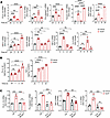 Figure 3
SP-NK1R signaling increases transcripts for proteins of the NLRP3 inflammasome and synthesis of pro–IL-1β and TNF-α in mouse keratinocytes. (A) Quantification by qRT-PCR of transcripts for proteins of the NLRP3 inflammasome in COCA cells at indicated times following treatment with SarSP (10–7 M) or vehicle. (B) Detection of pro–IL-1β and TNF-α protein by ELISA in COCA cells at indicated times following treatment with SarSP (10–7 M) or vehicle. (C) Comparative analysis by qRT-PCR of pro–IL-1β, pro–caspase-1, TNF-α, and IL-6 transcripts in primary cultures of keratinocytes isolated from K14Cre/+ NK1RKO (K14-NK1RKO) or littermate (Litt) control mice. In A and B, each circle represents 1 culture of COCA cells (n = 3 and n = 4, respectively). In C, each circle represents a primary keratinocyte culture from 1 mouse (n = 3). Means ± 1 SD were analyzed by 1-way ANOVA and post hoc Student-Newman-Keuls test; *P < 0.05, **P < 0.01, ***P < 0.001, ****P < 0.0001.
Following skin penetration with haptens, IL-1β and TNF-α released locally initiate the inflammatory response required for the sensitization of ACD (10–14, 68). Thus, we verified that the increased expression of IL-1β and TNF-α mRNA detected by qRT-PCR in COCA cells treated with SarSP correlated with higher content of pro–IL-1β protein in cell lysates and of secreted TNF-α in culture supernatants of COCA cells treated with SarSP under the same conditions (Figure 3B).
Next, we analyzed whether the increased expression of mRNA for components of the NLRP3 inflammasome and for proinflammatory cytokines detected in keratinocytes treated with SarSP was mediated through signaling via NK1R. Analysis by qRT-PCR demonstrated that cultures of skin keratinocytes from K14Cre/+ NK1RKO mice, unlike those from littermate (Creneg) controls, failed to upregulate the mRNA of pro–IL-1β, pro–caspase-1, TNF-α, and IL-6 in response to SarSP (Figure 3C). Thus, signaling of keratinocytes via the SP-NK1R axis augments expression of mRNA encoding proteins involved in priming and assembly of the NLRP3 inflammasome required for synthesis and secretion of IL-1β and increases synthesis and release of TNF-α — the 2 cytokines that initiate the cutaneous innate immune response during hapten sensitization (14, 69).
The SP-NK1R axis induces pro–IL-1β and TNF-α in keratinocytes via different intracellular signaling pathways. TNF-α primes and sustains the expression of the NLRP3 inflammasome under aseptic conditions and is an essential cytokine for the induction of ACD (63–67). Thus, we investigated whether pro–IL-1β in keratinocytes treated with SarSP is induced by the neuropeptide or indirectly through release of TNF-α. Comparative qRT-PCR analysis in cultures of keratinocytes obtained from WT or from TNF-αKO mice treated with SarSP or vehicle demonstrated that TNF-α is necessary to increase the expression of pro–caspase-1 and NLRP3 mRNA, but not pro–IL-1β mRNA (Figure 4A). Thus, SP-NK1R signaling of keratinocytes augments expression of pro–IL-1β in a TNF-α–independent fashion but increases pro–caspase-1 and NLRP3 transcripts in a TNF-α–dependent manner.
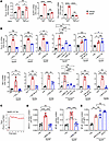 Figure 4
SarSP stimulates intracellular signaling pathways involved in synthesis of pro–IL-1β and TNF-α. (A) Quantification by qRT-PCR of transcripts for pro–IL-1β, NLRP3, and pro–caspase-1 in primary cultures of keratinocytes isolated from WT or TNF-αKO mice. (B) Analysis by qRT-PCR of the intracellular signaling pathways involved in the increase of pro–IL-1β and TNF-α transcripts in COCA cells treated with SarSP (10–7 M) alone or with intracellular pathway inhibitors or their vehicles (Veh). (C) FACS analysis of Ca2+ flux induced by SarSP (10–7 M) in COCA cells. A representative experiment of 3 is shown. (D) Quantification by ELISA-based assay of NFATc1 and NF-κB nuclear translocation in COCA cells treated with SarSP (10–7 M) alone or with the mouse NK1R-specific antagonist RP67,580 (10–5 M) or vehicle. (E) Quantification by ELISA-based assay of NF-κB nuclear translocation in COCA cells treated with SarSP (10–7 M) alone or with intracellular (BAPTA-AM) or extracellular (EGTA) Ca2+ chelators. In A, each circle represents a keratinocyte primary culture from 1 mouse (n = 3). In B, D, and E, each circle represents a single culture of COCA cells (B, n = 3; D, n = 4; E, n = 3). Means ± 1 SD were analyzed by 1-way ANOVA and post hoc Student-Newman-Keuls test; *P < 0.05, **P < 0.01, ***P < 0.001, ****P < 0.0001.
Next, we analyzed the intracellular pathways triggered by the SP-NK1R axis associated with the increase of expression of pro–IL-1β and TNF-α mRNA in keratinocytes. We investigated this in cultures of COCA cells treated with SarSP together with inhibitors of Gαq/11 (YM-254890), NF-κB (JSH-23), TNF-dependent NF-κB translocation (BAY11-7082), PI3K (CGS15943), calcium-dependent NFAT (VIVIT peptide), and the MAPK proteins JNKII (SP600125), p38 (SB203580), and ERK (PD98059). The qRT-PCR analysis showed that increase of TNF-α and pro–IL-1β transcripts by SarSP required signaling via Gαq/11, NF-κB, PI3K, and the MAPK proteins JNKII, p38, and ERK. Whereas increase of TNF-α transcripts depended on NFAT and TNF-dependent NF-κB translocation signaling, these pathways were not required for the increase of pro–IL-1β transcripts (Figure 4B).
Signaling via the NK1R induces intracellular Ca2+ increase in excitable and non-excitable cells, an effect that is needed for dephosphorylation and nuclear translocation of NFAT but is not necessarily required for NF-κB activation by SP (70–77). We confirmed in COCA cells that SarSP induced Ca2+ flux assessed by a ratiometric assay by FACS (Figure 4C), and nuclear translocation of NFAT and NF-κB, the latter inhibited by the mouse NK1R-specific antagonist RP67,580 (Figure 4D). However, SarSP stimulation of COCA cells in the presence of extracellular (EGTA) or intracellular (BAPTA-AM) Ca2+ chelators did not affect NF-κB nuclear translocation (Figure 4E).
In summary, these data confirm that SP-NK1R signaling in keratinocytes promotes per se the activation of key intracellular pathways, resulting in the synthesis of pro–IL-1β and TNF-α. In addition, the results show that SarSP increases the expression of pro–caspase-1 and NLRP3 transcripts in a TNF-α–dependent fashion.
DNCB induces pro–IL-1β but not TNF-α in mouse and human keratinocytes. The data to this point explain, at least in part, the molecular mechanisms by which the SP-NK1R axis promotes per se the proinflammatory function of keratinocytes, leading to the synthesis of pro–IL-1β and TNF-α, independently from each other. In addition, the data show that SarSP induces Ca2+ influx in mouse keratinocytes required for the NFATc1-dependent increase of TNF-α transcripts, and these effects were not necessary for the induction of pro–IL-1β transcripts. However, these findings do not elucidate the contribution of SP-NK1R signaling in the proinflammatory effect that DNCB exerts on keratinocytes during skin sensitization. Thus, we compared the proinflammatory effects that SarSP or DNCB exerts in COCA cells. DNCB (10 μM) increased NK1R expression as early as 2 hours after exposure of COCA cells (Figure 5A), but it did not induce intracellular Ca2+ increase (Figure 5B). Like SarSP, DNCB triggered nuclear translocation of NF-κB (Figure 5, C and D) and augmented expression of pro–IL-1β transcripts, and protein in cell lysates (Figure 5, G and I). Unlike SarSP, DNCB did not cause nuclear translocation of NFATc1 (Figure 5, E and F), increase of TNF-α mRNA, or the release of TNF-α protein in keratinocyte culture supernatants (Figure 5, H and J).
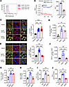 Figure 5
Synthesis of pro–IL-1β and TNF-α in mouse keratinocytes stimulated with DNCB or SarSP. (A) Expression (FACS) of the N-terminus of NK1R in COCA cells stimulated with DNCB (10 μM, 2 hours) or vehicle. (B) Ca2+ flux (FACS) induced by SarSP or DNCB in COCA cells. Left: one experiment of 3. Right: Comparison of Ca2+ flux by calculation of the area under the curve (AUC) (with FlowJo). Each circle represents 1 cell culture (n = 3). (C and E) Left: Representative images obtained by confocal microscopy of NF-κB (C) or NFATc1 (E) nuclear translocation in COCA cells treated with SarSP, DNCB, or DNCB vehicle (Veh). Right: Quantification of NF-κB (C) and NFATc1 (E) nuclear translocation analyzed in the images obtained by confocal microscopy using ImageJ/Fiji software and expressed as MFI of nucleus/cytoplasm ratio quantified in 20 cells across 8 independent HPFs; original magnification, ×600 (n = 20). Scale bars: 5 μm. (D and F) NF-κB (D) or NFATc1 (F) nuclear translocation by ELISA-based assay. Each circle represents an independent culture (n = 3). (G–J) Quantification in COCA cells treated as in C of pro–IL-1β and TNF-α mRNA by qRT-PCR (G and H) and pro–IL-1β and TNF-α proteins in cell extracts (I) or culture supernatants (J). In B, D, and F–J, each circle represents 1 cell culture (B, D, and G–I, n = 3; F and J, n = 4). Means ± 1 SD were analyzed by 1-way ANOVA and post hoc Student-Newman-Keuls test; *P < 0.05, **P < 0.01, ***P < 0.001, ****P < 0.0001.
To assess the clinical translational relevance of our findings, we investigated in human keratinocytes to what extent the SP-NK1R axis plays a role in inducing the keratinocyte proinflammatory changes triggered by DNCB. For these experiments we stimulated HaCaT cells or primary keratinocytes from adult skin (HEKa cells) with SarSP or DNCB. Analysis of increase of transcripts for pro–IL-1β and TNF-α confirmed that stimulation of HaCaT cells with SarSP was optimal at a concentration of 10–7 M (Supplemental Figure 4A). Similarly to what we observed in mouse keratinocytes, stimulation of HaCaT cells with SarSP increased the transcripts for relevant proteins involved in the NLRP3 inflammasome, including pro–IL-1β, pro–IL-18, pro–caspase-1, NLRP3, and TRPV1, and for the proinflammatory cytokines TNF-α, IL-6, and IL-8 (Figure 6A).
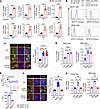 Figure 6
SP-NK1R signaling of human keratinocytes increases transcripts encoding proteins of the NLRP3 inflammasome and proinflammatory cytokines. (A) Quantification (by qRT-PCR) of transcripts for proteins of the NLRP3 inflammasome and proinflammatory cytokines in HaCaT cells treated with SarSP (10–7 M) at indicated time points. Each circle represents 1 keratinocyte culture (n = 4). (B) Left: Expression (by FACS) of functional NK1R assessed by the presence of the C-terminus in HaCaT and HEKa cells. Right: Expression of the N-terminus of NK1R in HaCaT and HEKa cells treated for 2 hours with DNCB (10 μM) or vehicle (Veh). One experiment of 3 is shown. (C and F) Left: Representative images by confocal microscopy of NF-κB (C) or NFATc1 (F) nuclear translocation in HaCaT cells treated with SarSP, DNCB, or DNCB vehicle. Right: Quantification of NF-κB (C) or NFATc1 (F) nuclear translocation analyzed with ImageJ/Fiji in images obtained by confocal microscopy, expressed as MFI of nucleus/cytoplasm ratio quantified in 20 cells across 8 independent HPFs; original magnification, ×600 (n = 20). Scale bars: 5 μm. (D and G) Quantification by qRT-PCR of transcripts for pro–IL-1β (D) and TNF-α (G) in HaCaT and HEKa cells treated with SarSP (10–7 M), DNCB (10 μM), or vehicle. Each circle represents 1 cell culture (n = 3). (E) Analysis of Ca2+ flux (FACS) induced by SarSP or DNCB in HaCaT cells. Top: One representative experiment of 3 is shown. Bottom: Comparison of Ca2+ flux by calculation of the area under the curve (AUC) (with FlowJo). Each circle represents 1 cell culture (n = 3). Means ± 1 SD were analyzed by 1-way ANOVA with post hoc Student-Newman-Keuls test; *P < 0.05, **P < 0.01, ***P < 0.001, ****P < 0.0001.
HaCaT and HEKa cells expressed the functional variant of the NK1R, which was increased by treatment with DNCB (10 μM) for 2 hours (Figure 6B). Both DNCB and SarSP induced nuclear translocation of NF-κB and increase of pro–IL-1β transcripts (Figure 6, C and D). However, SarSP but not DNCB induced intracellular Ca2+ increase and nuclear translocation of NFATc1 (Figure 6, E and F). Consequently, only SarSP upregulated TNF-α mRNA in HaCaT and HEKa cells (Figure 6G). Importantly, nuclear translocation of NFAT and NF-κB was inhibited by the human NK1R–specific antagonist L733,060 (Supplemental Figure 4B).
Administration of IL-1β plus TNF-α restores CHS in K14Cre/+ NK1RKO mice. The physiological relevance of the results obtained in vitro in mouse and human keratinocytes was assessed by comparison of the innate and adaptive immune responses to DNCB in Creneg littermate controls and K14Cre/+ NK1RKO mice sensitized only or sensitized and elicited with topical DNCB, the latter in the presence or absence of intradermal (i.d.) IL-1β, TNF-α, or both. Controls included mice treated topically with the DNCB vehicle plus PBS or with DNCB vehicle and IL-1β plus TNF-α (i.d.).
Analysis of ear and epidermal-dermal thickness and the intensity of skin-infiltrating leukocytes confirmed that lack of NK1R in keratinocytes of K14Cre/+ NK1RKO mice prevented the innate immune response to topical DNCB in comparison with littermate (Creneg) controls (Figure 7, A and B). The innate immune response to DNCB in K14Cre/+ NK1RKO mice was partially but significantly increased by i.d. injection of IL-1β or TNF-α in the sensitization site immediately after DNCB application, and it was fully restored and comparable to that of littermate controls following co-administration of both cytokines (Figure 7, A and B, and Supplemental Figure 5, A and B). As expected, i.d. administration of IL-1β and TNF-α in the absence of DNCB induced skin inflammation characterized by edema and inflammatory leukocyte infiltrate that peaked 24 hours after treatment, and these effects were significantly lower than the ones induced by skin co-administration of DNCB plus both cytokines (Figure 7A and Supplemental Figure 5, A and B).
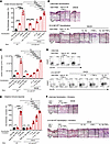 Figure 7
TNF-α and IL-1β restore the innate and adaptive immune response of DNCB CHS in K14Cre/+ NK1RKO mice. (A) Innate immune response by percentage of ear thickness 24 hours after sensitization of K14Cre/+ NK1RKO (K14-NK1RKO) mice treated on the right ear with topical DNCB alone or with IL-1β, TNF-α, or both (i.d.). Creneg littermates (Litt) were treated with topical DNCB alone. Controls included mice treated with topical vehicle (Veh) plus PBS (i.d.), or topical vehicle and IL-1β plus TNF-α (i.d.). Each circle represents 1 mouse (n = 5). (B) Microscopic images of mouse ears shown in A illustrating epidermal-dermal thickness (vertical lines) and skin-infiltrating leukocytes. Scale bars: 50 μm. (C and D) Quantification (FACS) of skin-migratory dendritic cells (smiDCs) in skin-draining lymph nodes (sdLNs) 72 hours after sensitization as in A. Each circle represents smiDC number per individual sdLN (n = 3). (D) Representative dot plots of the smiDCs in sdLNs. (E) Effector immune response by percentage of ear thickness in K14-NK1RKO mice sensitized in the abdominal skin with DNCB alone or with IL-1β, TNF-α, or both (i.d.) and elicited in the right ear 5 days later with topical DNCB alone or with IL-1β, TNF-α, or both (i.d.). Creneg littermates were sensitized and elicited with DNCB alone. Controls included mice sensitized and elicited with topical vehicle plus PBS (i.d.), or topical vehicle and IL-1β plus TNF-α (i.d.). Each circle represents 1 mouse (n = 5). (F) Microscopic images of the ears of mice in E 48 hours after elicitation, showing epidermal-dermal thickness (vertical lines) and skin-infiltrating leukocytes. Scale bars: 50 μm. (B and F) H&E; original magnification, ×200. Means ± 1 SD were analyzed by 1-way ANOVA and post hoc Student-Newman-Keuls test; **P < 0.01, ***P < 0.001, ****P < 0.0001.
Development of the adaptive immune response of ACD requires that skin-resident DCs migrate to the sdLNs to prime hapten-specific pathogenic T cells. Because (a) activation and traffic of skin-migratory DCs (smiDCs) depend on IL-1β and TNF-α released in the skin (10–12, 14, 15) and (b) expression of IL-1β and TNF-α mRNA in DNCB-sensitized skin is significantly lower in K14Cre/+ NK1RKO mice, we analyzed whether expression of NK1R in keratinocytes affects the number of smiDCs in sdLNs after DNCB sensitization. Flow cytometric analysis of sdLNs 72 hours after treatment showed that the number of total smiDCs identified by the phenotype CD45posCD11cposMHC-IIhi (Supplemental Figure 5C) was significantly decreased in K14Cre/+ NK1RKO mice compared with those in sdLNs of littermate controls (Figure 7C). Decrease of smiDCs in K14Cre/+ NK1RKO mice included epidermal Langerhans cells (LCs) (CD207posCD103negCD301bneg) and dermal type 1 cDCs (cDC1) (CD207posCD103posCD301bneg) and cDC2 (CD207negCD103negCD301bposCD11bhi) (Figure 7D and Supplemental Figure 5D). In the absence of DNCB, i.d. IL-1β and TNF-α increased the number of smiDCs in the sdLNs of K14Cre/+ NK1RKO mice. However, only DNCB plus both cytokines restored the numbers of smiDCs in sdLNs to those observed in Creneg littermate controls (Figure 7C and Supplemental Figure 5D). Likewise, the effector phase of CHS to DNCB was partially restored in K14Cre/+ NK1RKO mice injected i.d. with IL-1β or TNF-α at the DNCB sensitization and elicitation sites and was recovered to a level similar to that developed in littermate controls when both cytokines were co-administered with the hapten (Figure 7, E and F, and Supplemental Figure 5, E and F). As expected, i.d. administration of IL-1β and TNF-α in the absence of DNCB, analyzed 48 hours after elicitation, was not sufficient to stimulate the effector phase of the CHS (Figure 7, E and F, and Supplemental Figure 5, E and F).
In summary, lack of NK1R in keratinocytes is sufficient to prevent the skin innate and adaptive immune responses elicited by DNCB by a mechanism involving the synthesis and secretion of IL-1β and TNF-α.


